
Let’s just first try to understand the basic concept of kidney. You should know that a pair of bean-shaped structured organ which is known as kidneys are located just below and posterior to the liver in the peritoneal cavity. The suprarenal glands which are commonly known as adrenal glands place on top of each kidney. The prime operation of the kidney is to filter blood and purify it. The kidneys basically filter all the blood in the human body is multiple times a day. To perform such function these organs utilized up almost 25 percent of the oxygen inhaled through the lungs. Oxygen permits the kidney cells to efficiently produce chemical energy through aerobic respiration in the form of ATP. The filtrate coming out of the kidneys is known as urine.
On the outside, the kidneys are encircled by three layers. The renal fascia is the outermost layer which is a tough connective layer of tissue. Perirenal fat capsule is the second layer, that helps anchor the kidneys in place. Renal capsule is the third and innermost layer. On the inside, the kidney has three sections—an outer cortex, a medulla in the middle, and the renal pelvis in the region called the hilum of the kidney. The blood vessels and nerves come in and exit the kidney through the concave part of the bean-shape which is known as the hilum. Hilum is also the point of exit for the ureters. The occurrence of nephrons which is functional unit of the kidney makes the renal cortex granular. The medulla contains of several pyramidal tissue masses, called the renal pyramids. the blood vessels pass through the spaces known as renal columns which is placed in between the pyramids. Renal papillae which is the tips of the pyramids point toward the renal pelvis. There are around eight renal pyramids in each kidney. The renal pyramids that are present along with the adjoining cortical region are known as lobes of the kidney. In this case, the renal pelvis leads to the ureter and that too on the outside of the kidney. Coming to the inside of the kidney, the renal pelvis branches out into two or three extensions called the major calycesthat further subdivided into the minor calyces. The ureters are urine-bearing tubes that exit the kidney and empty into the urinary bladder.
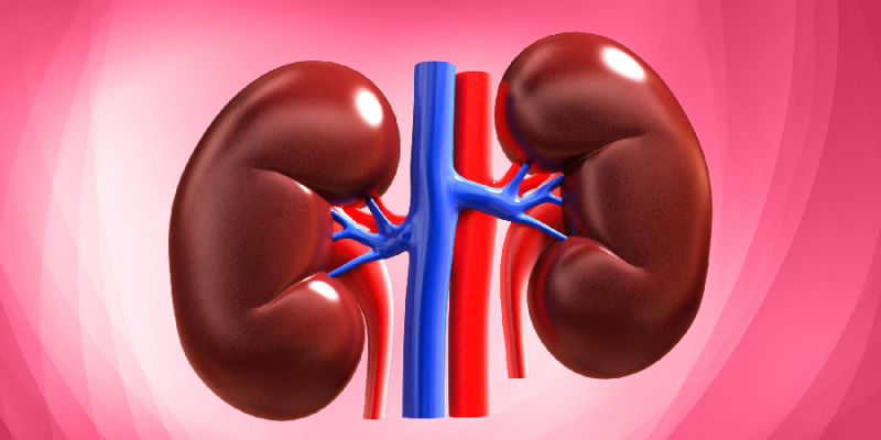
The network of blood vessels is a vital section of its structure and function of the kidney, as the main function of the kidney is to filters blood. The arteries, nerves, and veins which supply the kidney enter and exit at the renal hilum. Renal blood supply begins with the branching of the aorta into the renal arteries and ends with the exiting of the renal veins in order to join the inferior vena cava. The renal arteries branched into multiple segmental arteries upon entering the kidneys. Each segmental artery branched further into several interlobar arteries and enters the renal columns that provide the renal lobes. The interlobar arteries branched at the junction of the renal cortex and medulla to form the arcuate arteries. The arcuate “bow shaped” arteries happens to form arcs along the base of the medullary pyramids. Cortical radiate arteries, as the name indicates, radiate out from the arcuate arteries. The cortical radiate arteries subdivided into several afferent arterioles, and then pass in the capillaries supplying the nephrons. Veins happen to trace the path of the arteries and have similar names, except there are no segmental veins.
As I have mentioned previously, nephron is the most important functional unit of the kidney. Each kidney contains over 1 million nephrons which dot the renal cortex, giving it a granular appearance when sectioned sagittally. The nephrons can be classified into two types—cortical nephrons (85%), which are deep in the renal cortex, and juxtamedullary nephrons (15%), which lie in the renal cortex close to the renal medulla. A nephron comprises of three parts— a renal tubule, a renal corpuscle, as well as the associated capillary network, which originates from the cortical radiate arteries. The corpuscle gives rise to the proximal convoluted tubule that passes to the radiate part of cortex. The capillary network which originates from the renal arteries provides the nephron with blood that required to be filtered.


Comments